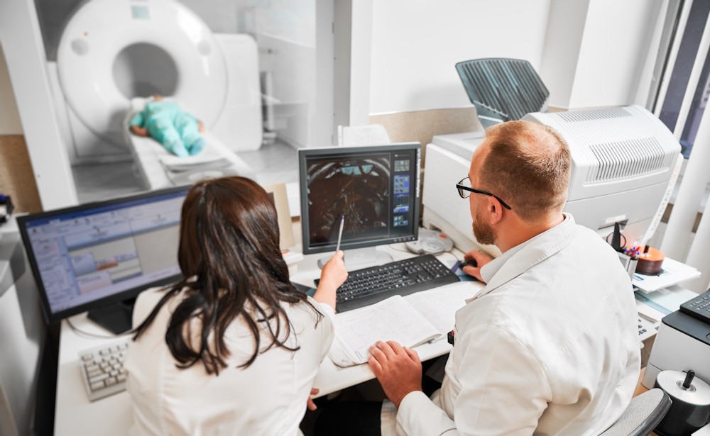
Diagnostic Tests Used by Orthopedic Specialists
The field of advanced orthopedics is no stranger to innovation. For example, arthroscopic surgery has proven to be a suitable alternative to open surgery, affording patients a far less invasive option that promotes shorter recovery times.
Another thing orthopedic specialists commonly do is provide diagnoses. To do that, they must perform an exam and look inside the body to see the affected joint. Although diagnostic imaging has been around for some time now, it remains one of the cornerstones of advanced orthopedics when determining the root cause of an orthopedic issue.
Orthopedic specialists generally rely on three types of diagnostic imaging tests:
- Computed tomography (CT) scans
- Magnetic resonance imaging (MRI)
- X-rays
Each modality brings unique strengths to the table, making them indispensable in different healthcare scenarios. In this blog, we’ll examine the distinctive features of MRIs, CT scans, and X-rays, exploring the specific scenarios they are used.
X-Rays – What Are They and When Are They Used?
X-ray technology has been around since the late 1800s, making it one of the oldest imaging methods in advanced orthopedics. Using electromagnetic energy beams and external radiation, X-rays can produce images of the body, organs, bones, joints, and tissue. X-rays are especially valuable to orthopedic specialists because they emphasize bone and joint structure, making it easy to identify abnormalities and injuries.
X-rays are typically used in the following healthcare scenarios:
- Screenings for cancer, arthritis, and other diseases that affect the musculoskeletal system.
- To identify structural issues in the joints, bones, or soft tissue.
- To identify foreign bodies or bone fragments.
- To identify the root cause of swelling, pain, or lack of mobility.
- To check for bone breaks and fractures.
The Application of CT Scans in Advanced Orthopedics
CT scans are an elevated form of diagnostic imaging used in advanced orthopedics. They offer a three-dimensional internal view of the body by combining X-ray technology with computer processing, allowing orthopedic specialists to see detailed images of bones, soft tissues, organs, muscles, and blood vessels.
Physicians rely on CT scans for a more detailed evaluation, especially when assessing complex fractures, soft tissue injuries, and conditions affecting bones and surrounding structures. Using a contrast agent – a substance that can be injected or taken orally – physicians can get an even more detailed look at the area in question.
CT scans are typically used for:
- Trauma assessment, such as injuries, complex fractures, and sports-related joint issues.
- Diagnosing soft tissue damage.
- Identifying abdominal issues and internal injuries.
- Examining small bones, blood vessels, and other parts of the anatomy that require more detail.
- Examining tumors and foreign bodies.
What Is an MRI and When Is It Used?
Magnetic resonance imaging (MRI) is the final diagnostic test we’ll examine. As the name implies, it employs powerful magnets and radio waves to create detailed images, particularly of soft tissues like muscles, ligaments, and cartilage, which is what makes it so useful in the field of advanced orthopedics.
Unlike X-rays and CT scans, MRIs do not use ionizing radiation. Patients can eat, drink, and take their medications as they normally would without impacting the results of their MRI. Although MRIs provide the most detailed internal view of the three diagnostic imaging tests, it is also the most challenging to administer. Patients must lie still within a tube-like enclosure for 30 to 60 minutes.
MRIs are used in numerous medical scenarios, including:
- Blood vessel and heart issues, including aneurysms.
- Ligament, tendon, and cartilage damage that impacts joint function.
- Inner ear and eye issues.
- Stroke and brain injuries.
- Cancer that affects the organs or bones.
- The diagnosis of multiple sclerosis (MS).
The Importance of Collaborative Decision-Making
In the realm of diagnostic imaging, the choice among X-rays, CT scans, and MRIs is a collaborative decision made by orthopedic specialists based on the specific clinical questions at hand. This collaboration ensures that the selected imaging aligns with diagnostic goals.
In many cases, the results of diagnostic imaging inform the patient’s treatment options. For example, MRI results that confirm a shoulder labrum tear mean that you need treatment from an orthopedic surgeon near you. Imaging results that show a minor sprain, on the other hand, mean the patient will avoid surgery and can heal naturally using holistic methods.
Some medical scenarios fall in the middle, meaning an orthopedic specialist and a surgeon will weigh the pros and cons of different treatment options while factoring in the needs of the patient. Undergoing surgery is often the quickest and most efficient way to address an orthopedic issue, and some patients are willing to endure it for the sake of their long-term mobility and function.
Advanced orthopedics affords physicians multiple ways of examining and treating injuries and medical conditions that affect bone and joint health. Patients can also consult with their orthopedic specialist about advanced pain management and treatments that accelerate healing, such as orthobiologics.
Consult with Physicians Who Lead the Field of Advanced Orthopedics
X-rays, CT scans, and MRIs each play a vital role in determining the root cause of musculoskeletal issues. The strategic use of these imaging modalities ensures a nuanced and comprehensive approach to diagnosis and treatment. By understanding the strengths and applications of each tool, patients have more insight as to what their orthopedic specialist can see once the scan is complete.
Those who are currently dealing with pain or mobility issues may benefit from having the area examined by an orthopedic specialist. You can kick things off by finding a practice near you that specializes in advanced orthopedics. During your initial appointment, your physician will likely perform at least one diagnostic imaging scan to get a better view of the area.
After making a diagnosis, your physician will explain your treatment options. They may also make a referral to an orthopedic surgeon near you for conditions that require more aggressive forms of treatment.

